 OVERVIEW
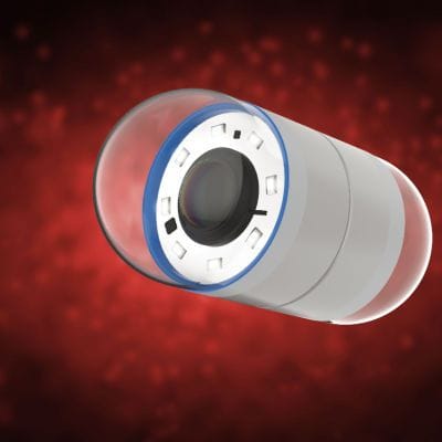
Capsule endoscopy is a procedure that makes use of a wireless camera in a capsule.
This type of endoscopy allows your gastroenterologist to get a live look at your small intestine (duodenum, jejunum, and ileum), which is the middle portion of the digestive tract. Video images of your gastrointestinal tract are recorded on an external device which can be viewed to detect and help diagnose or assess gastrointestinal conditions such as:
- Crohn's disease
- Ulcers, polyps and tumors of the small intestine
- Sources of bleeding from the small intestine
- Celiac disease
How does a Capsule Endoscopy Work?
A capsule endoscopy consists of a tiny wireless camera that's put into a capsule that the patient swallows. Prior to your exam, your doctor may have you drink some laxatives to clean out your digestive system in order to get higher-quality pictures.
The camera takes thousands of pictures as it makes its way down your digestive tract—the camera will take pictures for approximately eight hours, during which a recording device is worn on your waist records and stores the pictures.
Once complete, the capsule exits your body in your stool. Your doctor will then evaluate the pictures—which are combined together to form a video—and look for evidence of gastrointestinal disease.
Capsule Endoscopies are Safe When Performed by a Board-Certified Gastroenterologist
While capsule endoscopies are safe for most patients, they should be avoided if:
- You have a small intestine obstruction
- Your Crohn's disease has caused a narrowing in the small intestine
- You have a pacemaker or defibrillator
Complications of capsule endoscopy are rare but include the capsule getting stuck in a narrow portion of the digestive tract. This is typically caused by inflammation, scarring from prior surgery, or a tumor. If you have abdominal pain, bloating, nausea, vomiting, or fever after capsule endoscopy, let your doctor know immediately.
Disclaimer:
The information on this website is provided for educational and information purposes only and is not medical advice. Always consult with a licensed medical provider and follow their recommendations regardless of what you read on this website. If you think you are having a medical emergency, dial 911 or go to the nearest emergency room. Links to other third-party websites are provided for your convenience only. If you decide to access any of the third-party websites, you do so entirely at your own risk and subject to the terms of use for those websites. Neither FLINT GASTROENTEROLOGY ASSOCIATES, PC, nor any contributor to this website, makes any representation, express or implied, regarding the information provided on this website or any information you may access on a third-party website using a link. Use of this website does not establish a doctor-patient relationship. If you would like to request an appointment with a health care provider, please call our office at (810) 603-8415.

 OVERVIEW

